Three questions regarding the measurements by the SCT:
We are generating csa.csv files using “sct_process_segmentation”. Can you please advice which of the columns are showing the spinal cord volume? Or what can be the equation to measure this?
We are generating lesionseg_analyzis Excel files from Axial and Sagital images with an equal number of lesions, but the measurements are very different in volume - can you share, based on your experience the differences between the two?
Is there a way to combine Sagital and Axial MRI images to improve the results? I tried converting Dicom to NII but the result was two files instead of a combined one (using dcim2niix).
We are generating csa.csv files using “sct_process_segmentation”. Can you please advice which of the columns are showing the spinal cord volume? Or what can be the equation to measure this?
If you output the slicewise csv (i.e., one CSA per slice), you can compute the cord volume by summing the column “MEAN(area)” across the desired number of slices, and then multiply the result by the slice thickness.
We are generating lesionseg_analyzis Excel files from Axial and Sagital images with an equal number of lesions, but the measurements are very different in volume - can you share, based on your experience the differences between the two?
The difference is related to the precision of your measure. With thick sagittal and axial slices, the delineation of the lesion is hampered by partial volume effect, so you will inevitably experience this loss of precision. One way to handle this is to compute lesion volume in both images and then average your results. For more details, see this article, which points to open-source code that deals with this situation.
Is there a way to combine Sagital and Axial MRI images to improve the results? I tried converting Dicom to NII but the result was two files instead of a combined one (using dcim2niix).
Thank you so much for your answers, they were really helpful!
We are working now to produce segmentation of the white & gray matter with SCT 4.0.0.
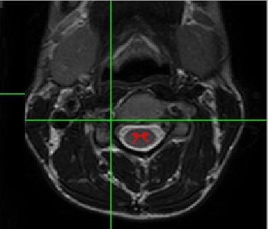
We were able to segment the Gray Matter using: sct_deepseg_gm. Please see the following screenshot:
Is it possible to measure (to Excel) the Gray Matter volume/area? We couldn’t use the tools mentioned in 2016 article since they have been changed.
In addition, we were unable to segment the White Matter - we couldn’t find an appropriate tool for that. Can you share the tools used, if any, to segment the White Matter as well?
Dear Julien
I hope you are well in these catastrophic times…
We have been working on our data of spinal cord MRI with the program. We found that if we repeat the analysis again we get different numbers.
Please advice
Dear Julien
It seems that the reason for the different results were related the implementation of white and grey matter measurements which we added.
When we re-tested the data results were the same!
Thanks for that.
The SCT version we use is 4.1.1
It’s been an overwhelming year, I hope everything is fine with you and your family!
We have another question regarding the CSA: We are measuring the spinal-cord volume of the same patient across different dates and MRI scanners. Is there a way to normalize the slices/measurements that we get from the SCT scripts, so we will measure from the same level of the spine?
At the moment, when we run the script, we sometime have 16 slices and sometimes we have 11, so the total volume is different.
When you specify the vertebral level with sct_process_segmentation, it will automatically select the levels you would like to compute CSA on. That way you are certain to measure CSA from the same level across subjects. For example, the following command:
Will compute an average CSA across levels C2, C3 and C4. The reported slices in the .csv file will necessary be different across subjects, because some subjects have longer vertebrae than others. For example in this subject, C2 → C4 is covered by slices 142 → 206, which is indicated in the output file csa.csv:
While that “total volume” does vary across individuals, the average CSA is normalized by the numbers of slices. For example, let’s imagine two participants with the exact same spinal cord size, and the size does not vary along the superior-inferior axis. In subject 1, the CSA is computed across 11 slices, while in subject 2 it is computed across 16 slices. The two CSAs will be exactly the same.