we are trying to find an optimal setup for the anatomical acquisitions of spinal MR. Referring to the course material for the SCT workshop in London and the SCT paper (2017), we are a little bit confused about the scanning parameters for the provided T2*-weighted images. We already tried several protocols provided by Siemens and also checked those you uploaded on OSF. Still, we couldn’t find a setting that provides such a nice representation of the spinal grey matter.
We would really appreciate any advice, which sequence and protocol parameters were applied for this scan or how to obtain a similar representation of the spinal cord.
Thank you for reaching out. Have you followed the SOP, which notably describes how to position the slice, the saturation band and the shim box? Those three parameters make quite a large impact on the resulting image quality. Finally, this sequence is quite sensitive to subject motion (including swallowing).
What scanner and coil are you using?
if you can share an example data, maybe i can assess the source of the problem (type of artifact, sensitivity profile, slice positioning etc.).
I have a similar inquiry as @cmelzer. I am trying to obtain the GM and WM in the spinal cord region that innervates to the legs (lower thoracic). Based on your recommendations I have tried increased averaging, anterior coil, and acquisition parameters proposed by [Yiannakas et al. PLOS One 2014] . I am using a Siemens Prisma scanner as well and known of the methods used have been consistent. Do you have any parameters that would yield the most consistent results?
Dear @jcohenadad
We want to add t2* series in order to get better segmentation of the gray matter. I don’t see t2* protocol in the link you share here. Should we follow the ME-GRE protocol? Is it t2*? Also what is the recommended TR/TE values?
Thanks a lot,
Juda
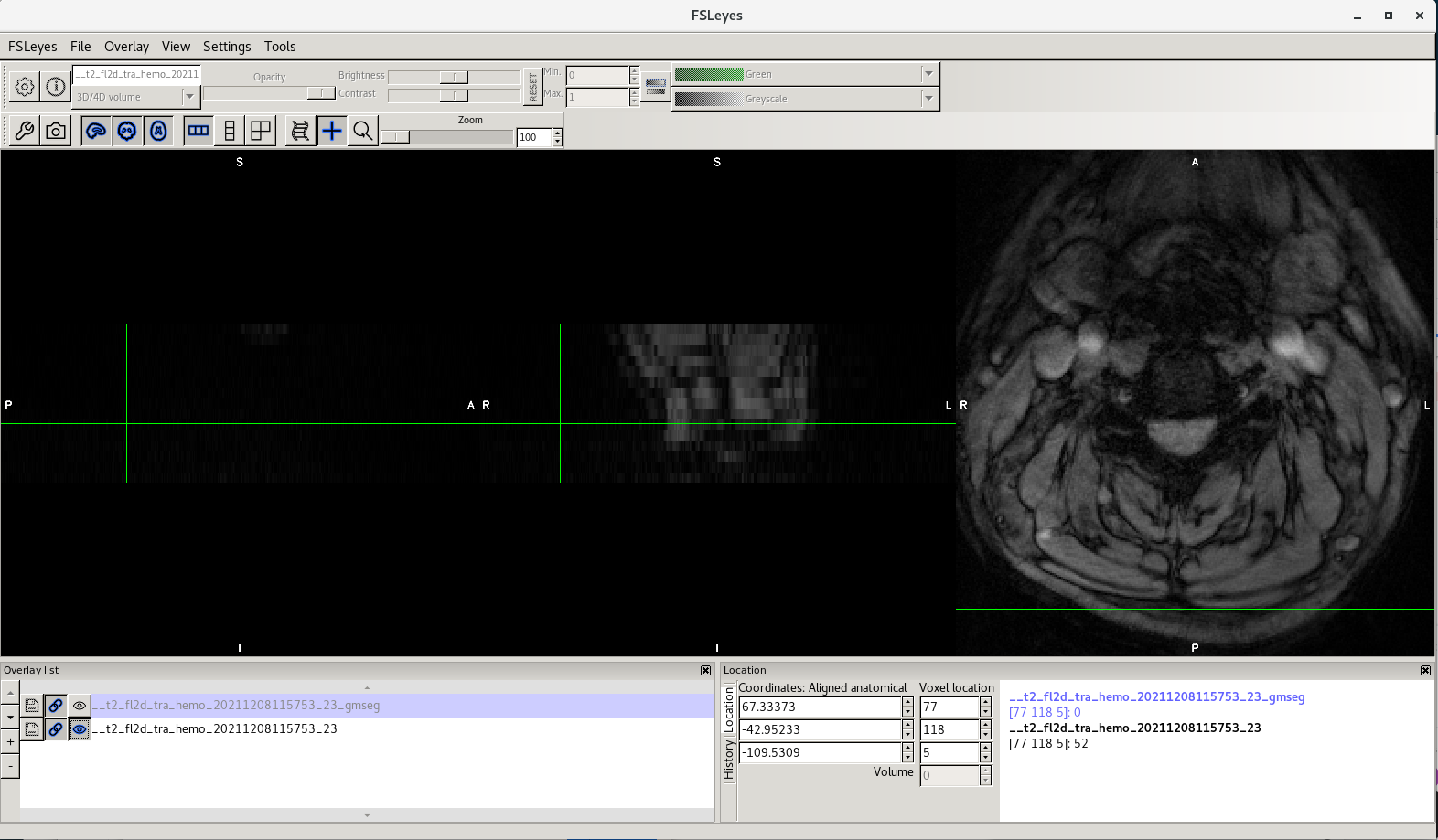
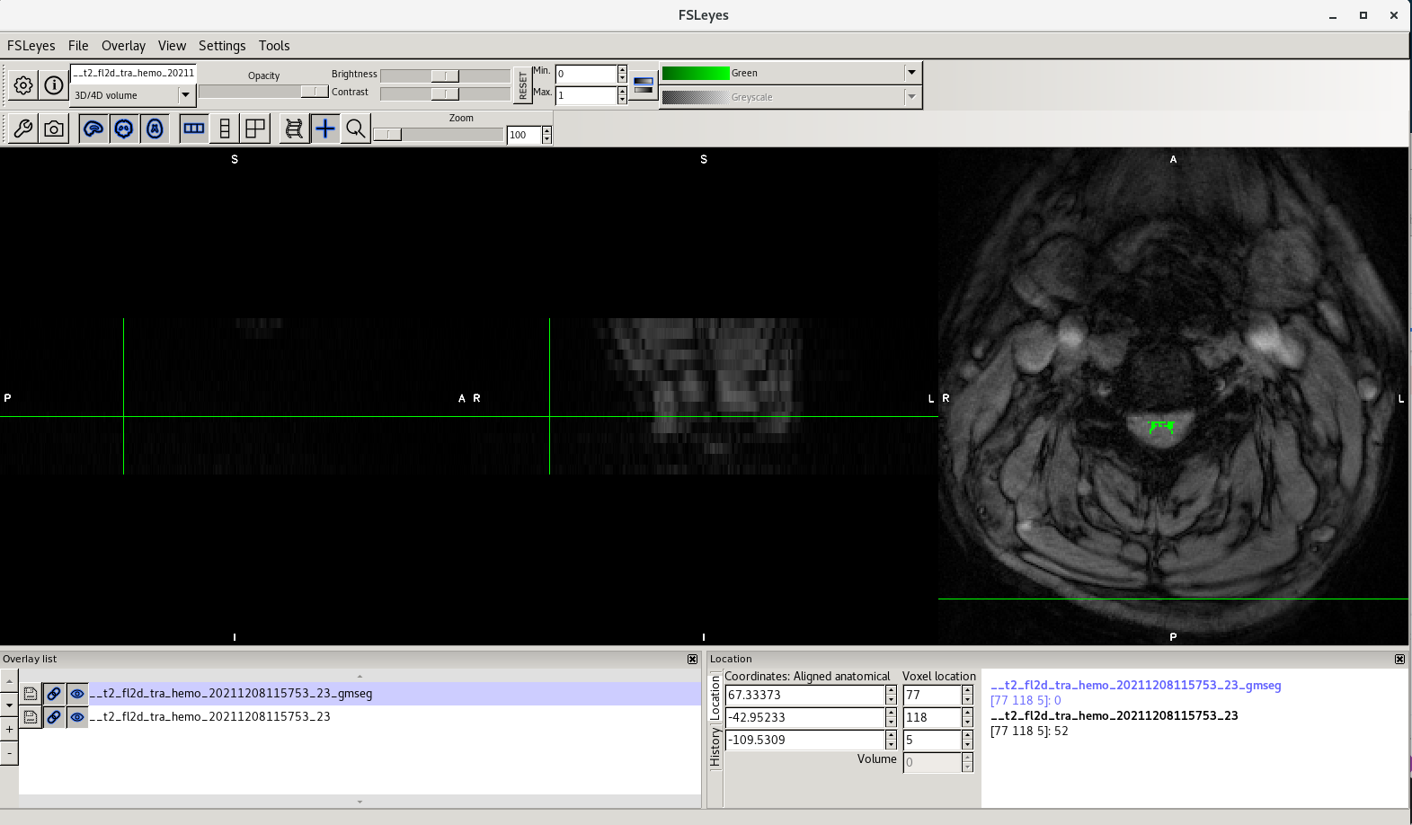
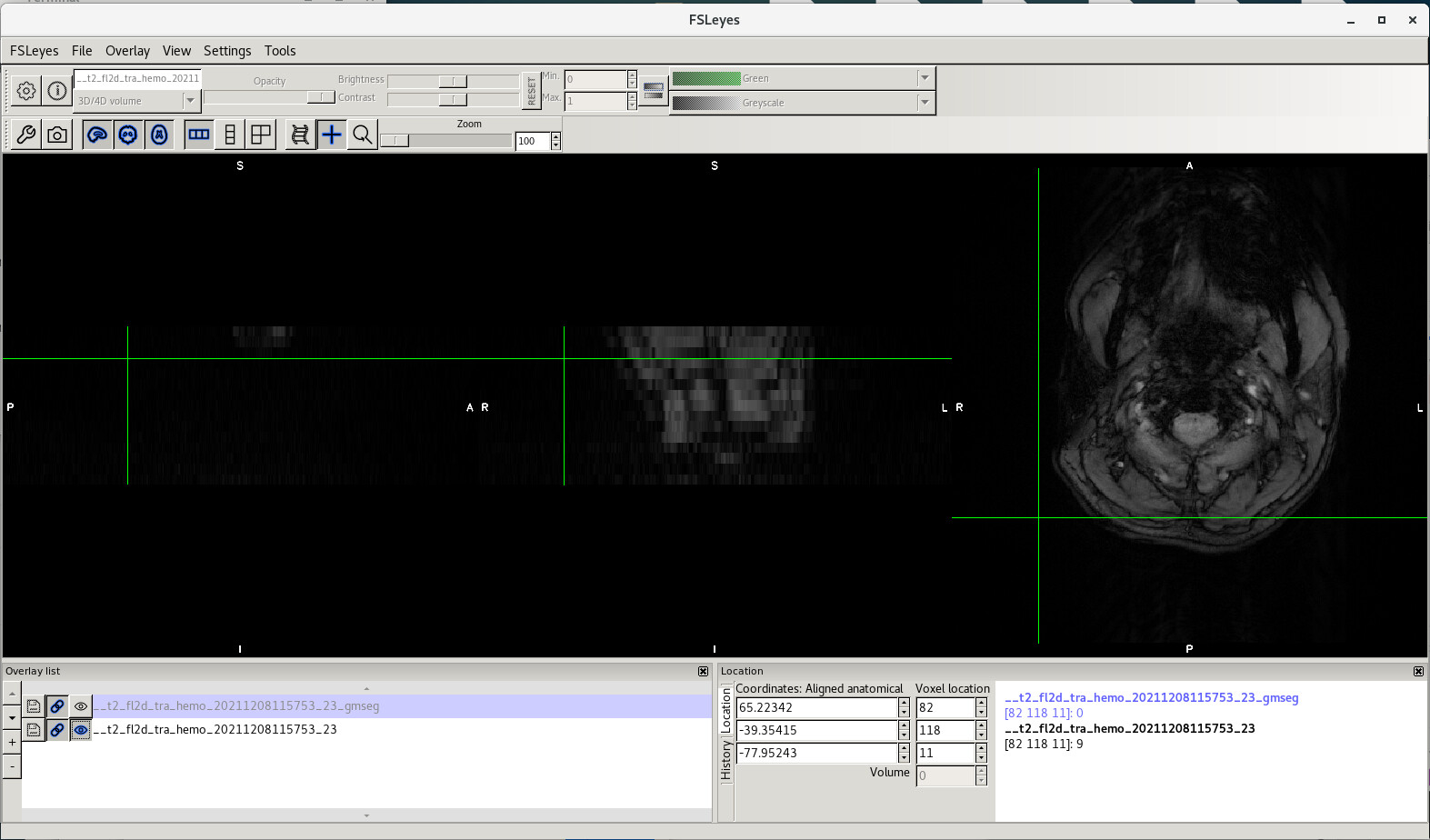
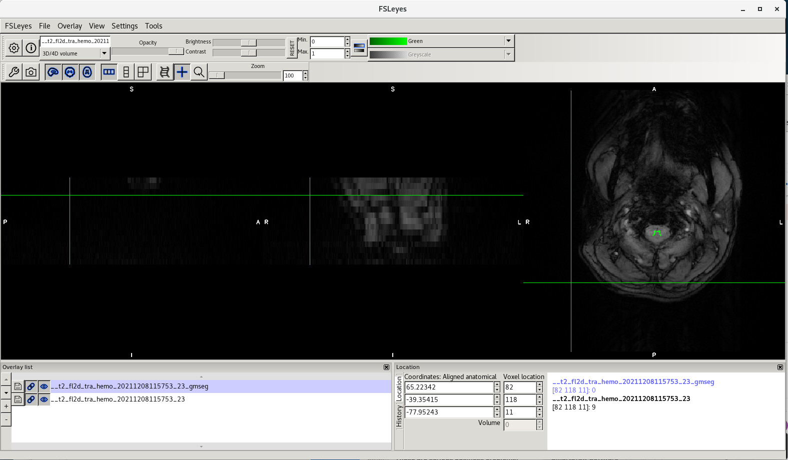
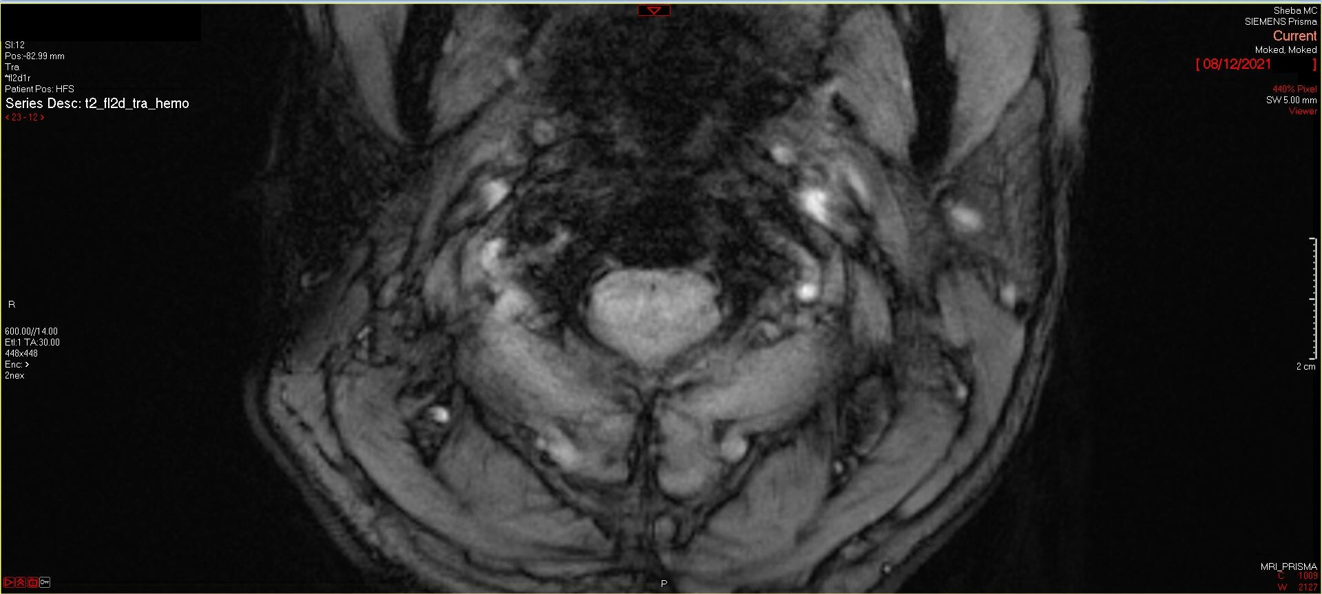
We tried to use the suggested parameters for the gray matter segmentation on one patient and we’re planning to continue with the study. When lookin at the results there is some slices in which we can see some of the gray matter, but in most of them we can’t see if clearly. Is it because of patient motion? the scanner is not fit for the task? or it should be like this? Is the result reliable enough to get good results of gray matter CSA when the segmentation seems ok, even if we can’t really see the gray matter in the original image?
Indeed, this ME-GRE sequence is highly sensitive to patient motion, which can produce ghosting and limits the ability to distinguish the gray matter. If you don’t see the gray matter with your naked eyes, there is no point in running the automatic segmentation. It won’t produce reliable results.
It does take some experience to acquire high quality ME-GRE data in the spinal cord. Few considerations include: proper patient positioning, careful slice orientation and shimming. This article details some of these tips, with example images of “good” acquisitions.