I would like to start by sincerely thanking all the developers and contributors who make this software available. It has provided invaluable insight into my daughter’s clinical case. I also wanted to share how I used SCT as a small case study, with the hope of demonstrating to patients, caregivers, and clinicians that this software is both accessible and powerful. Even for someone with limited knowledge of MRI technology or specialized software, it offers a meaningful way to extract deeper, quantitative insight from imaging data.
I believe my experience illustrates how approachable and impactful this tool can be. I am confident that radiologists will rely on software like SCT extensively in the future—it’s only a matter of time.
The MRI reports said nothing had changed—but everything was changing.
The Clinical Challenge
My daughter was diagnosed more than a year ago with a diffuse, expansile lesion in her spinal cord, likely a grade 2 spinal astrocytoma. Following an extensive work-up, two major research hospitals agreed to proceed with treatment without a biopsy. Over the course of her diagnosis and treatment, she has undergone eight MRIs—four during diagnosis and four afterward, as she progressed through chemoradiation and then chemotherapy.
Throughout this time, the MRI reports consistently described the lesion as “essentially unchanged.” However, clinically, her symptoms told a very different story: her deficits worsened prior to treatment and began improving afterward.
Taking Matters Into My Own Hands
About halfway through this process, I started taking a closer look at the MRIs using Horos and educating myself on the underlying technology. As a retired engineer, I found the challenge both engaging and compelling. Given that the lesion was described as “expansile,” I wondered whether I could quantify its progression.
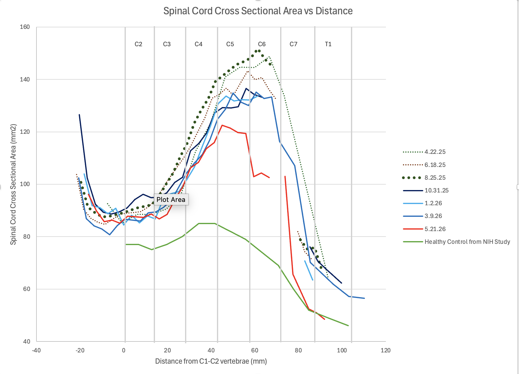
I began measuring the cross-sectional area of the spinal cord at different points, plotting these values along the length of the spine, and comparing them over time. To do this, I carefully adjusted each MRI sequence to help ensure they were comparable. I manually traced the spinal cord in axial views so Horos could calculate the area, corrected for differences in imaging angles, and aligned all data to consistent anatomical reference points.
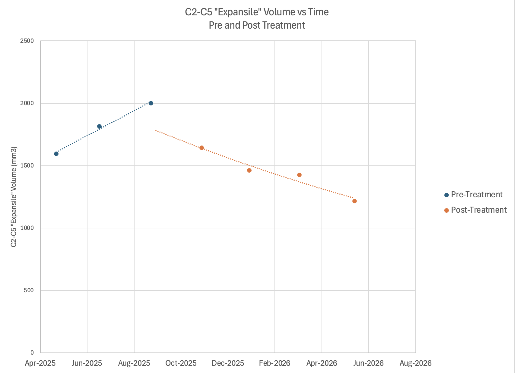
I incorporated a dataset of healthy control measurements along the spinal cord that I found in PubMed. Integrating the difference between my daughter’s data and these healthy controls, became a measure of cord expansion.
The results were striking. The total expansion increased steadily before treatment—matching the worsening of her symptoms—and decreased after treatment began, aligning with her clinical improvement.
For the first time, we could see the disease progression reflected clearly in the data.
A Breakthrough: Discovering SCT
Not long after, I discovered SCT through the research paper that had the same healthy control dataset. Encouraged by my son—who works in computer science—we decided to explore it further.
What followed was nothing short of remarkable.
I have no background in Python, and it had been years since I worked with advanced software tools like SCT. I expected a steep learning curve and wasn’t even sure SCT would outperform my manual workflow. Still, I was eager for an unbiased analysis, especially given my personal connection to the data.
Using AI + SCT Together
My son loaded the SCT software on our computer and introduced the software to Codex (ChatGPT’s coding module), along with the SCT training materials. Codex independently explored the SCT documentation and quickly learned its capabilities.
What happened next felt like having the software developer sitting right beside me.
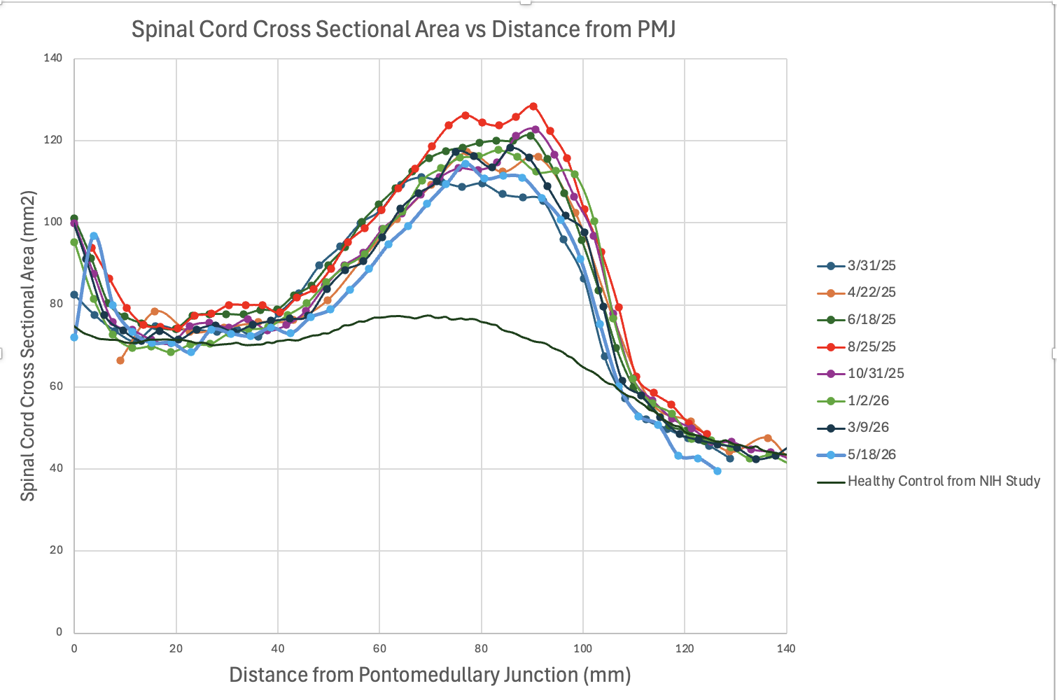
I simply described the analysis I wanted in plain language. Codex created all the terminal commands to have the MRIs analyzed by SCT, focusing on T2 sagittal and axial views across all dates. It walked me through SCT’s quality control outputs, which made it easy to identify and correct errors early on.
We used the PMJ distance feature to align the data and fine-tuned the positioning to ensure the expansile regions lined up accurately. Then I asked to incorporate the PAM50 healthy control dataset and align it with my daughter’s data. Finally, I requested a calculation of total expansile volume based on the difference between my daughter’s data and healthy controls within a defined spinal region.
Validation and Results
The results were astonishing. The SCT-generated data closely matched my manual measurements.
The results matched my independent analysis almost exactly.
This validated my approach and confirmed that my earlier findings were not biased. It was both reassuring and exciting to see that a tool like SCT could produce such consistent and reliable results with far less manual effort.
Manual Method
SCT generated data
Manual Method
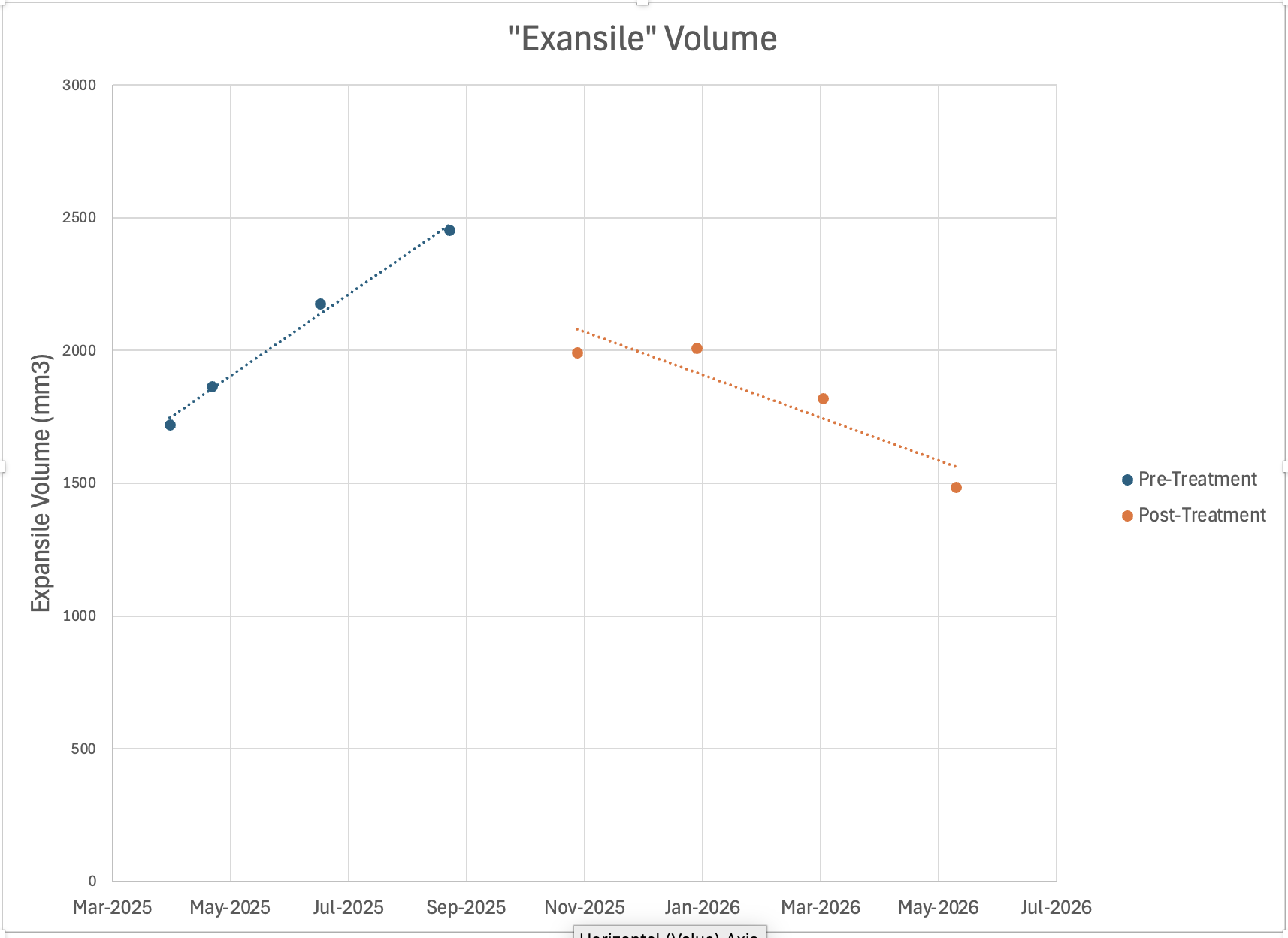
SCT generated data
What Surprised Me Most
There were several axial views that I had previously deemed too unclear to analyze confidently. In fact, I had abandoned the entire March 2025 sequence and much of the C6–C7 region of every other sequence because I did not trust the quality of the data.
SCT, however, was able to process all of them. The output demonstrated that it could extract meaningful insights even from images I considered unusable.
SCT was able to make sense of data I had completely written off.
Final Thoughts
Looking back, I only wish I had discovered SCT sooner. Not because my manual work wasn’t valuable, but because SCT made it faster, more consistent, and more objective.
My experience shows that tools like SCT can empower patients and caregivers to better understand complex medical data. With the addition of modern AI interfaces, even sophisticated analysis can become accessible through simple, plain-language interaction.
As these tools continue to evolve, they have the potential to change how MRI data is interpreted—not just in research settings, but in everyday clinical care. And perhaps most importantly, they can help bridge the gap between what imaging reports say and what patients are actually experiencing. This will also be incredibly useful for earlier detection of recurrence to allow for timely treatment.