dear jcohenadad, thank you for your help , I have got the result . but I have a question, in the past, I used the matlab to deal with the dti , however I think the output data are different from the sct . I could not understand the meaning of the output data from the sct. could you tell me what is the fa and md , rd value from the output data ?
dear jcohenadad , the problem is the difference between matlab and sct, I don’t know which row is the data I want. this data are output of the matlab .
the problem is the difference between matlab and sct
I don’t know which software you are using in Matlab to process your diffusion MRI data, therefore I don’t know what difference you are referring to. Could you please be more specific as to what exactly you would like to quantify and compare?
okay, thank you for your patience. I am really grateful.
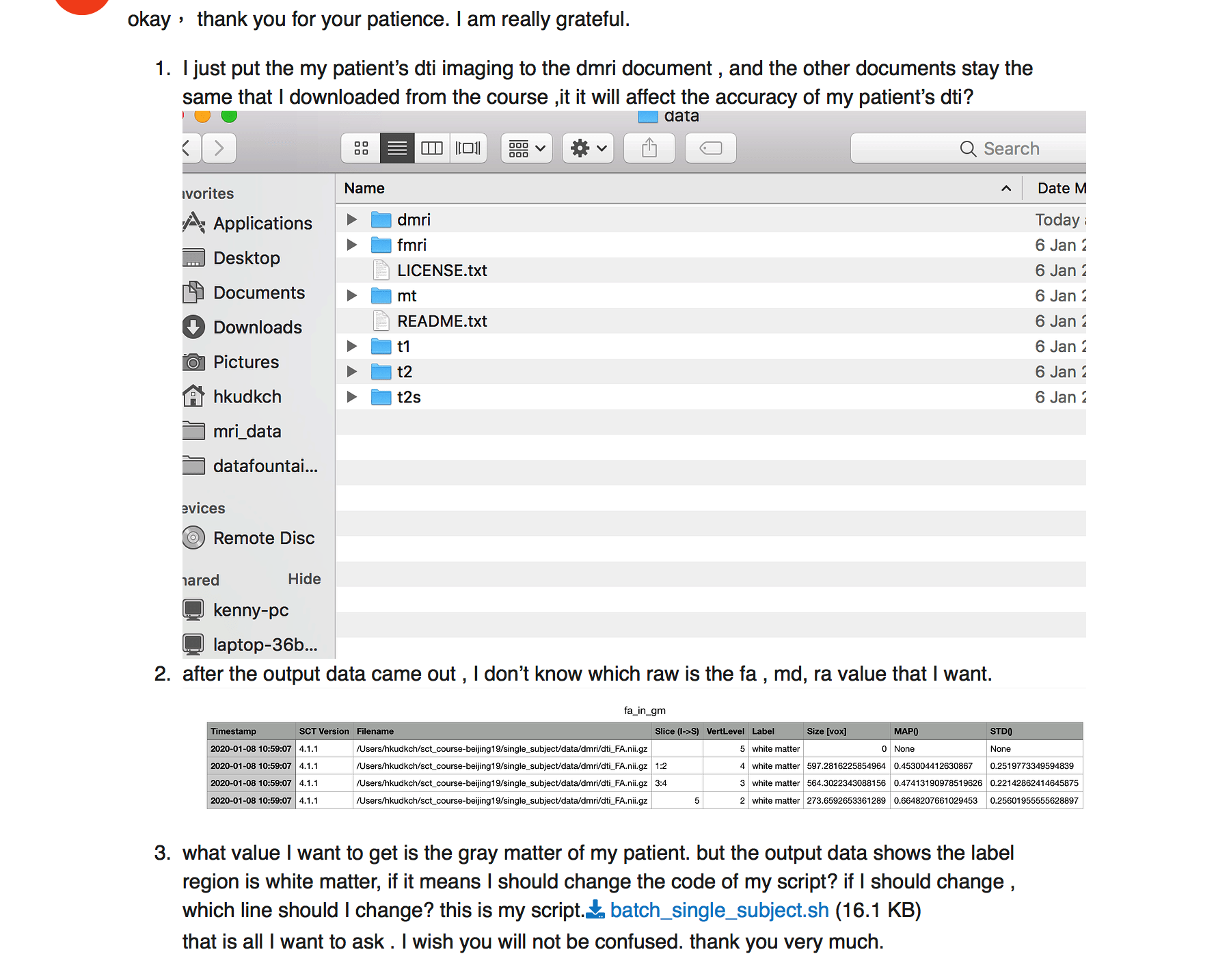
I just put the my patient’s dti imaging to the dmri document , and the other documents stay the same that I downloaded from the course ,it it will affect the accuracy of my patient’s dti?
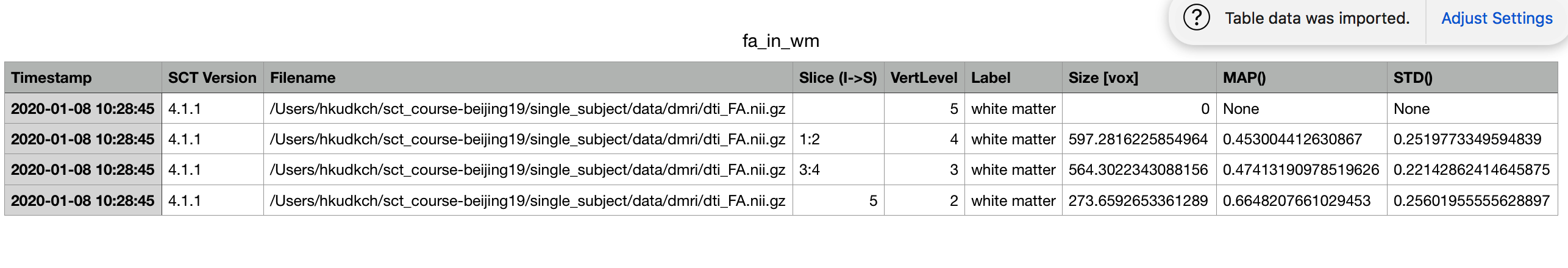
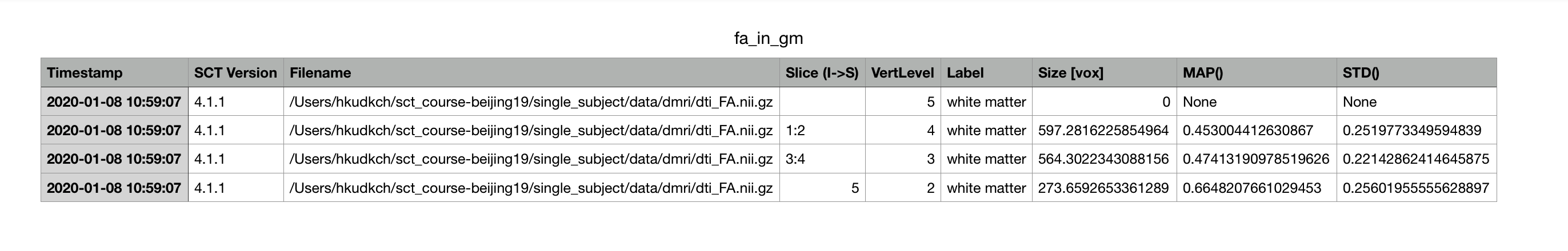
what value I want to get is the gray matter of my patient. but the output data shows the label region is white matter, if it means I should change the code of my script? if I should change , which line should I change? this is my script.batch_single_subject.sh (16.1 KB)
that is all I want to ask . I wish you will not be confused. thank you very much.
I’m sorry but I don’t understand your question. Could you please rephrase?
Each row corresponds to a vertebral level. Depending on your research project, you might have specific hypotheses related to the vertebral level. You could also output a single value averaged across multiple levels, but again, it depends on your research project.
Use flag -l 52 instead of -l 51 from the function sct_extract_metric. All that is explained in the SCT course material, which I encourage you to follow.
dear jcohenadad , I just put the my patient’s dti imaging to the dmri document , and the other documents stay the same that I downloaded from the course ,it it will affect the accuracy of my patient’s dti?
it means only the imaging in dmri document is belong to my patient, the imanging in other document don’t belong to my patient. if it affects the accuracy of my patient’s dti in dmri document.
thank you .
I want to explain , that I just put my patient’s dti imaging to the dmri document, and the other document is other patient’s(I don’t know who) imaging.( fmri, t1 ,t2 and so on). if I could get the right dti result of my patient?
my English is poor, please forgive me .
what do you mean by “I just put my patient’s dti imaging to the dmri document”. Did you mean folder? (instead of document)
if that’s what you meant, then you should not do that as the other images (t1, t2, etc.) are not from the same patient, and these data are used to process your dti data (e.g., warping field, etc.).
dear jcohenadad, thank you for your reply.
I think I have understood the sct.
1.I have the question. I have only one t1 imaging of c1-c7. however ,I have 3 t2 imaging (c1-c3,c3-c5,c5-c7),and 3 dti imaging (c1-c3,c3-c5,c5-c7). if I could put t1(c1-c7) in t1 folder, and t2(c1-c3) in t2 folder, dti(c1-c3) folder to calculate the dti (c1-c3) data ?
2. I think I have no the imaging of t2s ,what should I do? these are all imaging I have.
you don’t absolutely need the T1 or T2 scan to compute DTI data. You could also register the PAM50 to your DTI scans without it, if you know the vertebral level of your DTI scans.
Now, if you want to use an anatomical image, you can either use the T1 or the T2 image, depending which one has the best spatial resolution (especially axial resolution). All these aspects are explained in the SCT course.
Regarding the 2nd question: as mentioned above, you do not need to have the T2s data in order to quantify the DTI data.
thank you ,
sct_register_multimodal -i $SCT_DIR/data/PAM50/template/PAM50_t1.nii.gz -iseg $SCT_DIR/data/PAM50/ template/PAM50_cord.nii.gz -d dmri_crop_moco_dwi_mean.nii.gz -dseg dmri_crop_moco_dwi_mean_seg.nii.gz -param step=1,type=seg,algo=centermass:step=2,type=seg,algo=bsplinesyn,slicewise=1,iter=3 -initwarp …/ t2s/warp_template2t2s.nii.gz -initwarpinv …/t2s/warp_t2s2template.nii.gz -qc ~/qc_singleSubj
This script need the t1 and t2, could you tell me how to avoid this?
First of all, this syntax does not require a T1w or T2w image. It is using the template (PAM50_t1.nii.gz). Secondly, you can avoid relying on an initial warping field (here a T2*w-based field) by running sct_register_to_template directly on your DTI image, as I mentioned above. Examples are provided in the course material, or on the sct-pipeline repos.
Dear jcohenadad
When I put the dti imaging into the t1 folder and t2 folder to make a segementation. However when it came to this step sct_propseg -i t2.nii.gz -c t2 -qc ~/qc_singleSubj(I replace the t2 with dti. and I change the name of dti.nii to the t2. nii) Then it shows ERROR: your input image needs to be 3D in order to be segmented. What is the problem?
The problem is that you input a 4d image. You need to average your dmri series across time before inputting it into propseg, as exemplified in the SCT course.
At this point it will be easier if I just write the batch script for your analyses instead.
Just let me know what metrics you want, send me an example dataset and I’ll provide the script for you
TANG_WING_KUN_WIP_DTI_thk7_breath_1_5_1.zip (738.2 KB)
Dear jcohenadad , thank you very much.
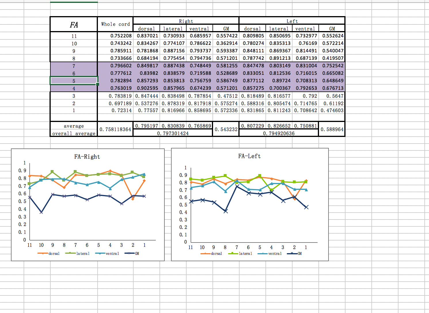
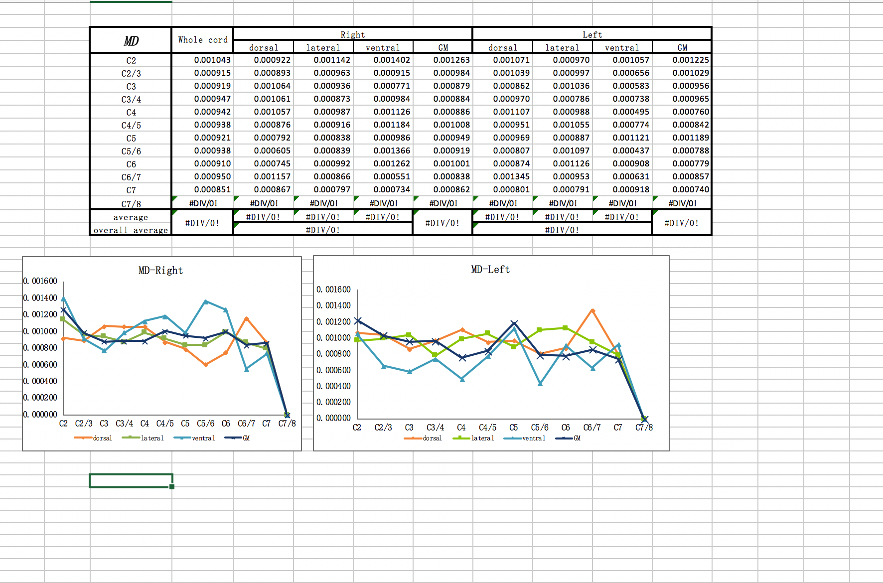
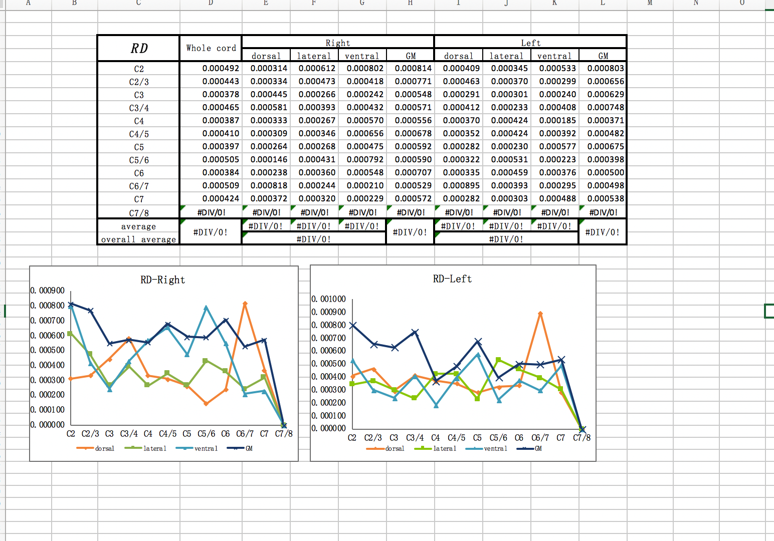
I want to get the patient’s gray matter‘s dti data , MD ,FA,RD value through this imaging I uploaded.
Thank you a lot!
Dear jcohenadad
I am sorry to bother you so long time , but I met too much problems before , what the data I want to get just like the picture I uploaded . these are the data of C1-C7 , the data I give you is C1-C3. but I think if I could deal with the DTI of C1-C3, it is enough for me.
Thank you.
hi,
sorry i am extremely busy these days. i was planning to work on your project probably sometimes next week. So, if I understand correctly: are you saying that you don’t need my help anymore?