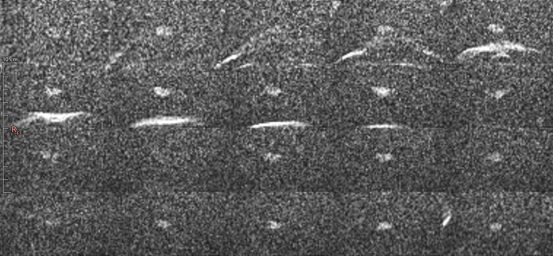
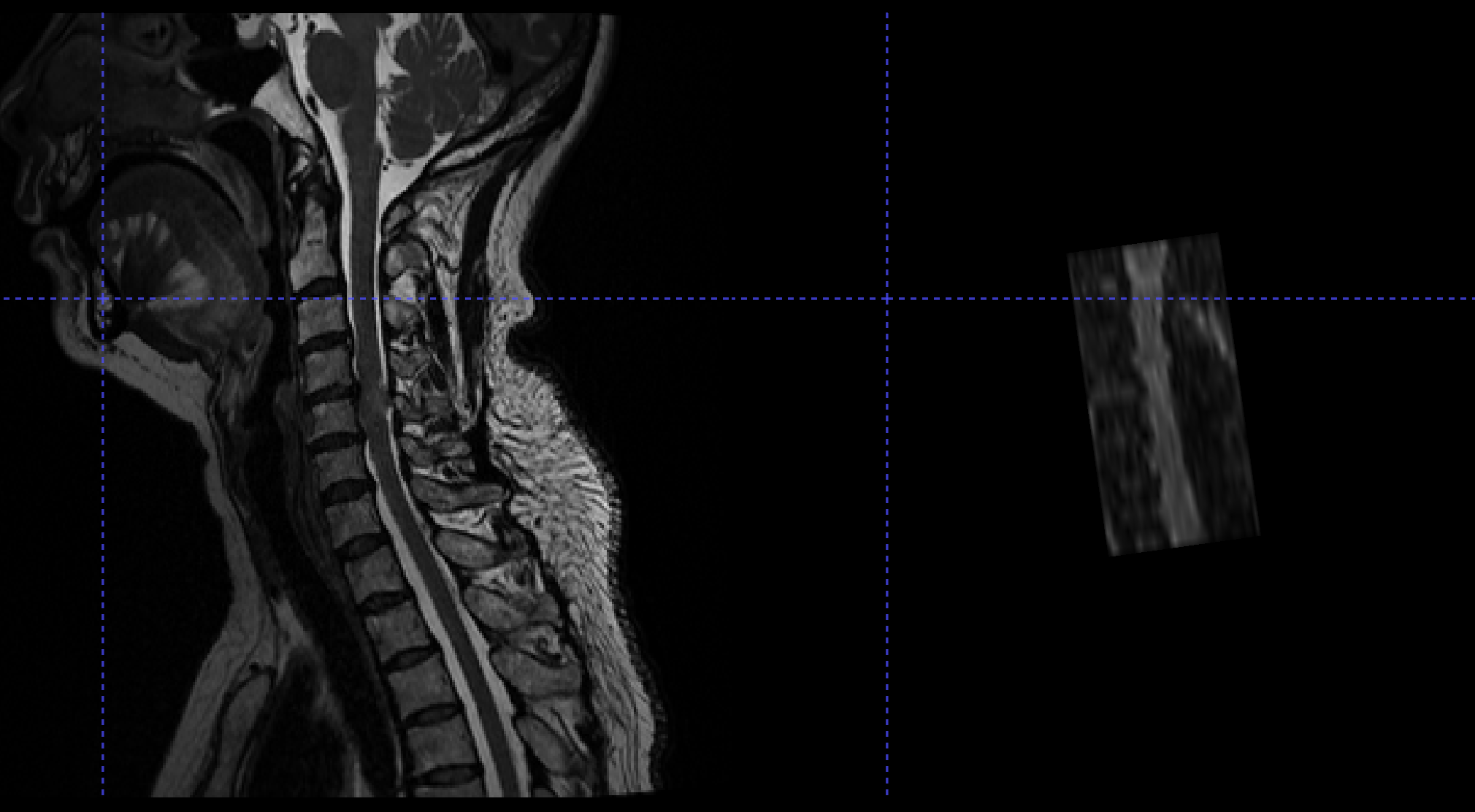
We have created a spine DWI protocol for United Imaging based on the generic protocols for Siemens. Oftentimes we encounter rainbow-shaped artifacts that could overlay the spinal cord. When reviewed on sagittal view, the artifacts seems to consistently be on the same level with the mandible. So we guess that those artifacts are caused by the jawbone. Any help is appreciated!
My deepest apologies for the late response. I am writing to let you know that @jcohenadad is currently out of office, and will return on August 28th.
I will check in with other members of the lab to see if they can provide insight into your question. But, otherwise, I thank you for patience until Julien returns.
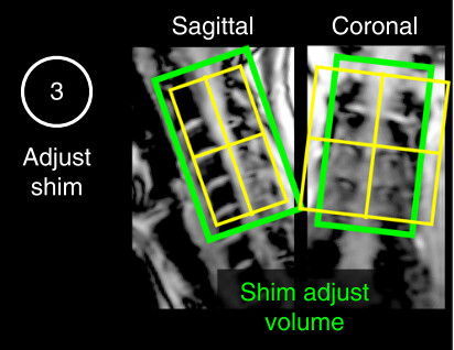
as Jan rightfully guessed, the rainbow-like artifact is caused by poorly suppressed fat due to insufficient shimming in that region. However, prescribing the shim region as shown in the picture above might not solve this, as the fat is typically outside of this green box— so in your case you might want to try increasing the coverage of the green box. Also note that the protocol was not validated on a United Imaging system, and I don’t know how the fat sat is implemented (pulse shape, offset frequency etc.) among other hardware and software possible differences.