Thank you so much to the SCT team for your previous assistance with the lumbar enlargement matching issue, which has now been fully resolved.
We are currently facing a new challenge related to gray matter segmentation . While we can successfully segment the gray matter in our data, we are looking for code or commands to further subdivide the segmented gray matter into specific regions (e.g., ventral,medial and dorsal regions) and calculate their respective areas. Could you provide guidance or tools to achieve this?
Furthermore, regarding the selection of white matter regions of interest (ROIs), the PPT mentions “Less than 2% error in ‘large’ tracts.” How is this “large” region defined? For example, is there a minimum number of pixels required to ensure more accurate DTI metric measurements?
I’m really looking forward to your help. Thank you.
Sunny
Dear Sunny,
Thank you for your follow-up question! We would be happy to help. ![]()
we are looking for code or commands to further subdivide the segmented gray matter into specific regions (e.g., ventral,medial and dorsal regions) and calculate their respective areas. Could you provide guidance or tools to achieve this?
We do have an Atlas-based analysis tutorial – the GM/WM atlas within the PAM50 template contains masks that span different sub-regions of the gray/white matter. Typically the atlas is used for metric extraction in those areas (i.e. DTI metrics), however here you are looking for area as well. I believe in this case you should be able to use sct_process_segmentation with specific masks.
Additionally, you may wish to use “GM-informed registration” along with sct_warp_template -a 1 to bring the PAM50 atlas to the space of the subject (while accounting for the shape of the gray matter). Then, you can compute the CSA of the atlas masks in the subject space.
Also, I notice you mention larger regions (ventral/media/dorsal) as opposed to individual tracts. To solve this problem, you can combine the sub-regions together. For an example of this, please refer to this page within the tutorial.
I am not quite sure what PPT you are referring to. But, in our Atlas tutorial, we have this section which explains the -method map approach:
The
mapmethod is the most robust to noise in small tracts. This was further validated using bootstrap simulations based on a synthetic MRI phantom. For more details, see [Lévy et al., Neuroimage 2015] (construction of the phantom, effect of noise, contrast) and [De Leener et al., Neuroimage 2017; Appendix] (effect of spatial resolution).
This may be what the 2% is referring to? But, I will double-check with my colleagues regarding the minimum size required for accurate measurements.
Kind regards,
Joshua
Dear Joshua
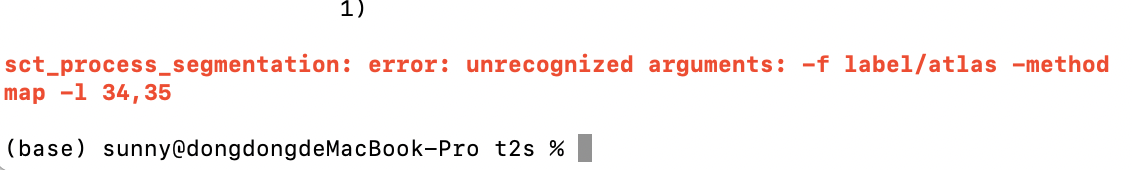
Thanks for your response!I am performing segmentation of gray matter subregions for calculating their areas. If using PAM50 template matching and atlas-based calculations, could the requirements for registration accuracy be exceptionally high due to the inherently small size of the gray matter areas? Additionally, I attempted to compute gray matter subregional areas using atlas registration encountered errors with the modified command–“ sct_process_segmentation -i t2s_gmseg.nii.gz -f label/atlas -method map -l 34,35 -o csa-gm1.csv “.
Sunny
Hi there,
I concur with @joshuacwnewton’s excellent suggestions. The very important thing, here, as already mentioned by Joshua, is that the analysis needs to be done in the subject’s space, otherwise you will end up calculating CSAs of the PAM50 atlas, instead of that from your specific participant.
Another, related, comment, is that by registering the PAM50 on your subject (using the SC and GM masks), the sub-regions will be quasi-linearly scaled. For example, if, let’s say, the SC, GM, dorsal columns of the PAM50 are respectively 60, 20 and 10 mm2, and if in your participant you calculate a 1.5x difference in CSA between the SC and the GM, the same (or very similar) ratio will apply to the dorsal column, giving you respectively 90, 30 and 15 mm2. My point is that you are not (and you cannot) directly measuring the CSA of sub-structure if you don’t see them on the MRI scans. You are only linearly scaling them with the SC and GM masks.
Based on what I wrote above, yes: the registration will be critical here. Also, you don’t want a registration method with too many degrees of freedom (such as SyN), which might yield a wrong representation of sub-structure CSA. I would go with more regularized approaches, eg BSplineSyN. But again, this is a delicate issue, and you want to make sure you have some sort of validation framework to make sure you’re doing things properly.
here, you want to input (with -i) the sub-region of interest, not the GM seg (see my comments above)–
Thank you very much for the previous guidance—I am currently working through it myself.
MD4-1_in_wm.csv (34.3 KB)
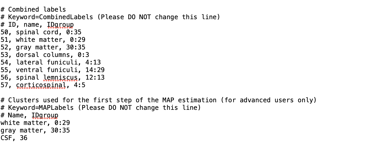
Furthermore, we are now working on add additional combinations of atlas.We have modified the info_label.txt under my SCT installation folder. the command of “the sct_warp_template” have been runned and the modified have been copied in my each subjects. However, there is no results about the additional combinations of atlas.
Hi
I have successfully run the code about the additional combinations of atlas and remain deeply grateful to your team.
Kind regards
Sunny
Indeed, the sct_warp_template simply copies the info_label.txt. The actual analysis on these combined tracts is performed later (using e.g. sct_extract_metric -l 57). I am very glad to hear that you solved the issue. ![]()
Thank you again for participating on the forum! We appreciate your questions and the discussion it provides. ![]()