I’m working with the neuronal segments in the lumbosacral region. So far I used my own segmentation based on the work of Frostell et al. I finally found the time to switch to your version introduced with 6.1 but realized that there is a major difference in the segmentation by more than a level at L3/L4.
I investigated a bit and I think I found the underlying issue; According to Frostell et al fig 1 (which you also show the PAM50 webpage) the caudal termination of the spinal cord is at intervertebral disk L1/L2. From table 5 and table 6 we get the information that the lumbosacral enlargement is at ‘Spinal Cord Segment’ L3 close to L4 or ‘Vertebral column segment’ T12. This matches with the alignment in fig 1.
If I calculate the LSE from the cord segmentation of the PAM50 the maximal value is around slice 172. This is however at the intervertebral disk T11/T12 in the middle of neuronal segment L2! This is the reason for my different segmentation.
So far I used T11/12 (intervertebral disc label 19) and L1/L2 (intervertebral disc label 21) as labels during registration for the LSE and caudal terminal of the spinal cord. Then I used the 2 points to calculate the neuronal segments with the information from Frostell et al in table 4. Using this registration with the newly provided neuronal segments puts information from the LSE which should be L3 to L4 into middle of L2.
I think there are two ways how I can handle this. I can either start using the point-wise vertebral body label from T12 as my (LSE) registration target and can then use the provided segmentation. This would however mean I register the native LSE to a point considerable below the PAM50 LSE and I’m not sure this is a god idea.
Or I keep the current registration process where I actually register native LSE to PAM50 LSE but then I have to use my original neuronal segmentation. I think this will produce better results however I don’t like the manual neuronal segmentation in regard to reproducibility and ability to compare.
Maybe I’m making an Error but I’m pretty sure the source of my problem is that the LSE of the PAM50 is not at diskT11/12 instead of midbody T12 as in Frostell et al. Any advice how to handle this situation?
I’m just writing to let you know that our team has read your post, and that we are currently discussing internally the best way to approach your issue. We will follow up after some further investigation.
Thank you for your excellent observations. We agree that, based on the PAM50 segmentation, the LSE is located slightly more rostral than what Frostell et al. predicts (ie: T11-T12 vs. mid T12). That being said, it should be noted that the segmentation of the PAM50 might be inaccurate, especially in the lumbar segments, where the contrast between the cord and the nerve rootlets is very low (ie: the CSA is possibly underestimated in the low lumbar spinal levels). So, it is possible, in fact, that the LSE is present at mid T12, but simply not reflected by the segmentation.
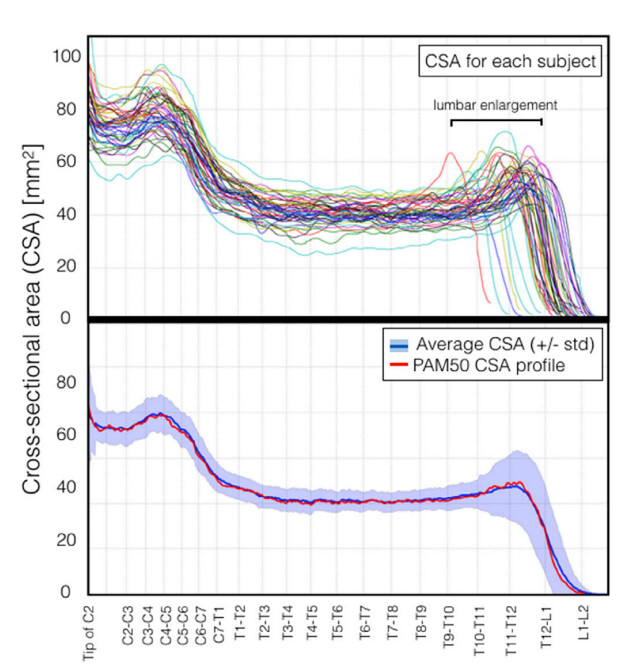
Another consideration: while Frostell et al. report the enlargement to be at L3-L4 spinal levels, that measurement itself is also relatively noisy, and possibly inaccurate. In fact, we did a similar analysis than the Frostell et al. in the PAM50 paper. When computing CSA along the spinal cord and displaying it across all the PAM50 subjects (with the cord aligned from the tip of C2 to L1-L2 vertebral levels), we notice a large variability of the LSE location (see top panel):
So, in light of these observations, instead of assuming that the LSE is perfectly located at mid. T12 across all subjects, we recommend to identify the LSE per subject, and use that label to do a single pointwise registration to the PAM50 template (using a label at mid T12 or at T11-T12, it doesn’t matter as long as it is consistent across all subjects). You could also consider using a second label, which would be the cauda equinea (value=60).
Also, we were a bit confused about your approach for registration. You mentioned you used Table 4 to get the neuronal segment from the vertebral levels, but Table 4 does not make any correspondance between the vertebral and the spinal segments. Maybe you meant Table 3?
To clarify this point: value=60 is a new feature that was added in SCT v6.1. For more details on the intended usage of this label, we have a tutorial focused on registration of the lumbar region here.
T12 across all subjects, we recommend to identify the LSE per subject, and use that label to do a single pointwise registration to the PAM50 template (using a label at mid T12 or at T11-T12, it doesn’t matter as long as it is consistent across all subjects)
That is what I’m doing. The question was which of these two you would suggest as targets. From your answer I assume using the the mid T12 label is better since it aligns with the new neuronal segment. My worry in this case was that the registration suffers from the fact that I’m registering my LSE and the corresponding binary mask to a point in the PAM50 and the cord mask which do not match. So above the label my mask becomes smaller while in the PAM50 it continues to become larger for another 30 slices/15mm. But this should not be a problem?
You could also consider using a second label, which would be the cauda equinea (value=60 ).
I’m aware, I used disc label 21 so far which is in the neighboring voxel. I intend to switch to the 60 one.
Also, we were a bit confused about your approach for registration. You mentioned you used Table 4 to get the neuronal segment from the vertebral levels, but Table 4 does not make any correspondance between the vertebral and the spinal segments. Maybe you meant Table 3?
Yes, my mistake. Table 3 was used to calculate the neuronal segments. But not in the registration process. Just to be sure the steps I’m doing listed:
I segment the spinal cord in my structural Image to create a binary mask.
From this mask I use the CSA to define the LSE and caudal termination slices and create a label file with disk labels 19 (LSE) and 21 (end).
(19 was chosen because it is next to the LSE slice of the PAM50 cord segmentation and 21 is going to be replaced with 60.)
I use image, mask and labels in the registration.
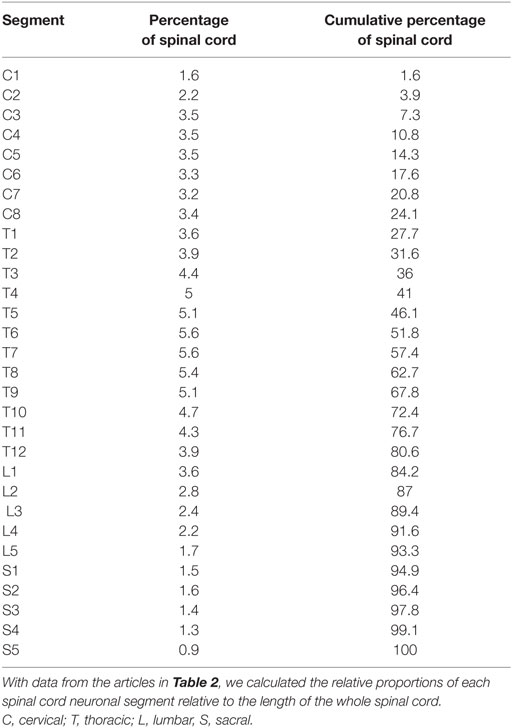
For the analysis I needed a neuronal segmentation of the cord. I did this with the information from Frostell et al. Table 5 and 3. Tab 5 gives me the information where the LSE has to be placed and tab 3 how the split the distances.
I now simply tried to switch my segmentation with the newly provided one and have the problem that my derived LSE target is in the middle of L2. Which raised the asked question if it is a good idea to use the T12 label as target for my LSE.
My understanding of what you are doing is slightly different. Based on what you wrote:
So far I used T11/12 (intervertebral disc label 19) and L1/L2 (intervertebral disc label 21) as labels during registration for the LSE and caudal terminal of the spinal cord.
I understand that you use vertebral-based markers to infer the location of LSE from Frostell’s Table 3, which is different than directly identifying LSE (eg: by looking at the maximum CSA per subject). This subtle difference has an implication on the subsequent processing. Indeed, if your approach is purely based on vertebral levels, then the corresponding spinal level do match between Frostell and PAM50, regardless of where the LSE is located. Or maybe I misunderstood your explanation? If you have your code somewhere we could look at it-- like pictures, code is sometimes worth thousands words.
Ah! So I did misunderstand then. You are indeed directly identifying the LSE for each subject. In that case, what you could do, if you wish to have the LSE exactly at L3-L4 as in Frostell et al., is to simply create an arbitrary label (eg: 59) on the PAM50_label_disc.nii.gz at mid-T12 that corresponds to the LSE, and use it for all your subjects alongside the already existing label 60 for the cauda equinea. I would not use 19 because this label already exists, and refers to T11-T12 disc.
That being said, I would like to reiterate that inferring the spinal levels based on the LSE is also problematic for at least two reasons:
Precise identification of the LSE is hard (because, in some subjects, the LSE can span several mm)
The correspondence between LSE and spinal levels in Frostell et al. is given for a group (not for individual subjects, for which that correspondance varies, as noted by the standard deviation).
So I think that combining two noisy measures, vs. only one based on intervertebral discs (but which are more clearly identifiable) is not necessarily going to provide the most accurate access to subject-wise spinal level. That being said, it would be great if you could compare both approaches in your fMRI study, and report which one yields more consistent results (if I was a reviewer I would definitely welcome such study ).