I have three DTI slabs covering the spinal cord from C1 to T6, with a voxel size of 1.3×1.3×5.0 mm. Do you recommend stitching them together before doing the analyses?
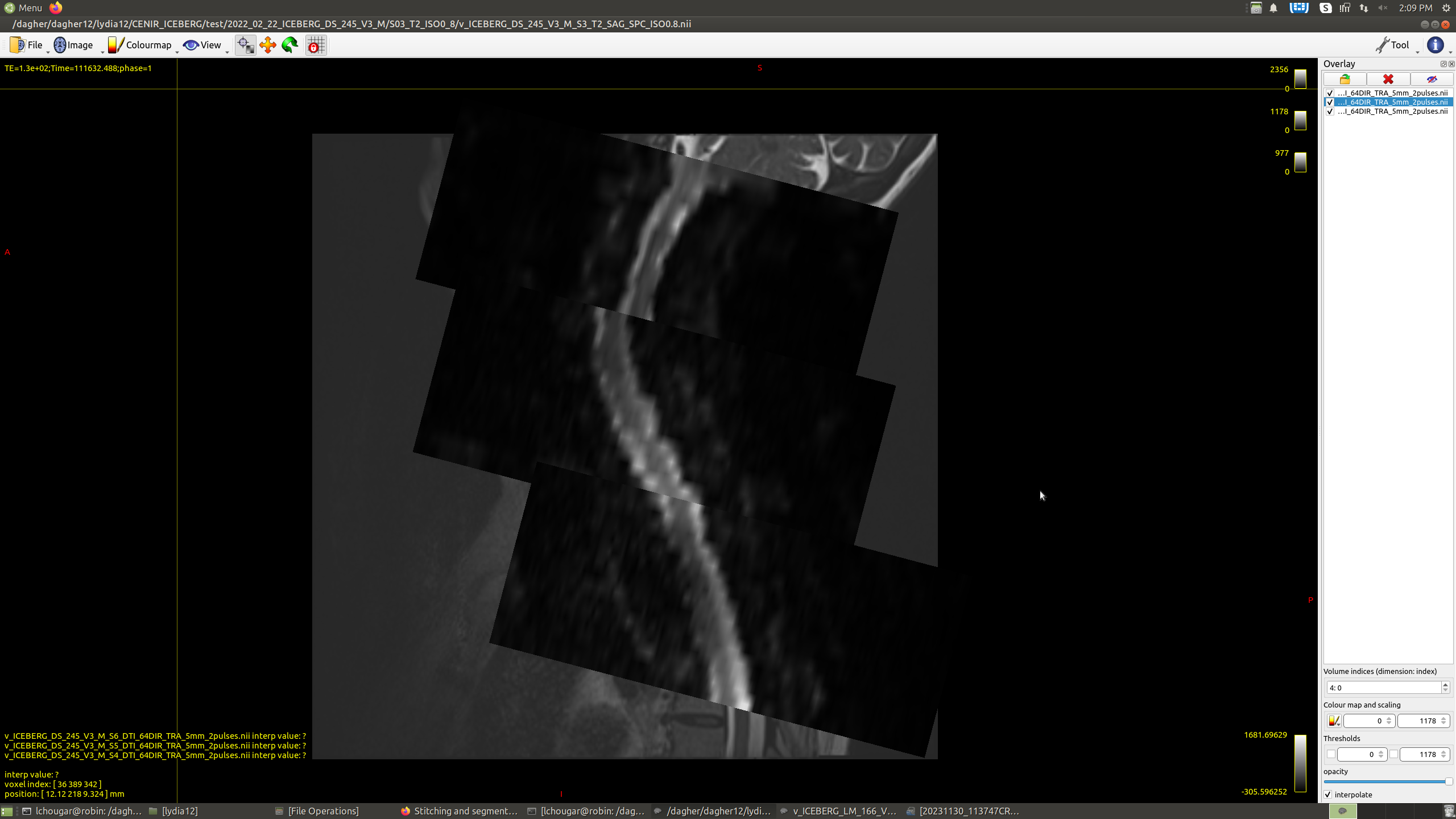
When I stitch them using sct_image -stitch, I lose the 4D information, and the slabs are slightly shifted. Is there a way to fix that?
Another question: in subjects with a marked cervical curvature, the segmentation fails at the level of the curvature. Is there a parameter in sct_deepseg_sc that I can adjust to make the segmentation more accurate?
Would you recommend systematically straightening the T2 images (using the sct_straighten_spinalcord command) before the segmentation?
I have three DTI slabs covering the spinal cord from C1 to T6, with a voxel size of 1.3×1.3×5.0 mm. Do you recommend stitching them together before doing the analyses?
I would not recommend stitching the raw (ie: DWI data), because the interpolation would impact noise distribution and model fitting. Instead, stitching should be done on the computed metrics (FA, etc.). The decision to stitch also depends on what the analysis will consist of. Eg: if the goal is to extract DTI metrics in specific tracts, then there is no need for stitching. Another thing to consider, is that the stitching algorithm assumes the slabs overlap sufficiently, and the structure overlaping the two slabs is perfectly registered. This assumptions is likely violated in the context of EPI scans, which are often hampered by susceptibility-related distortions.
Another question: in subjects with a marked cervical curvature, the segmentation fails at the level of the curvature. Is there a parameter in sct_deepseg_sc that I can adjust to make the segmentation more accurate?
The best way for us to help, would be if you could share a representative image privately. The optimization of parameters is impossible if we don’t see the images.
Would you recommend systematically straightening the T2 images (using the sct_straighten_spinalcord command) before the segmentation?
No, I would recommend against it. The curvature itself should not be the cause for a bad segmentation, because the default segmentation algorithm is 2D (ie: slicewise). However the curvature might cause particular undesirable effects in the image (eg: excessive CSF flow causing motion of the cord, causing image artifacts, causing the segmentation to fail). Again, without seeing the images, it is difficult for me to advise.
The raw DWI slabs don’t overlap perfectly in all subjects. Sometimes, they are adjacent but don’t overlap. In some subjects, they might be shifted as in the image. Then, it might be better not to stitch the FA, MD, …maps, right?
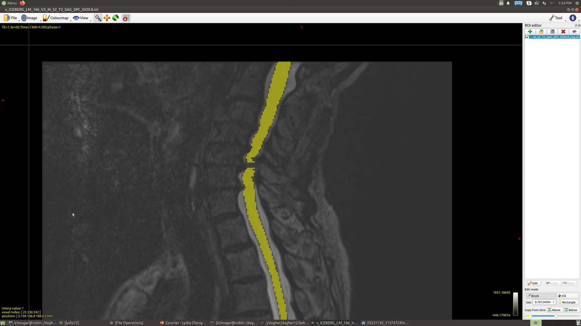
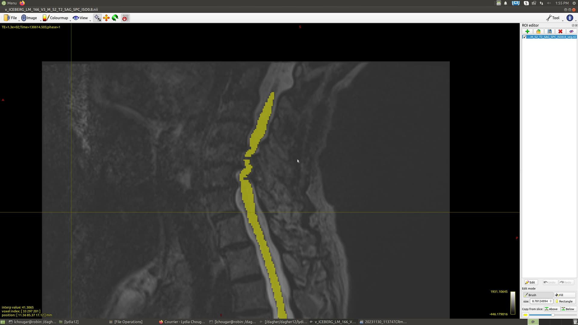
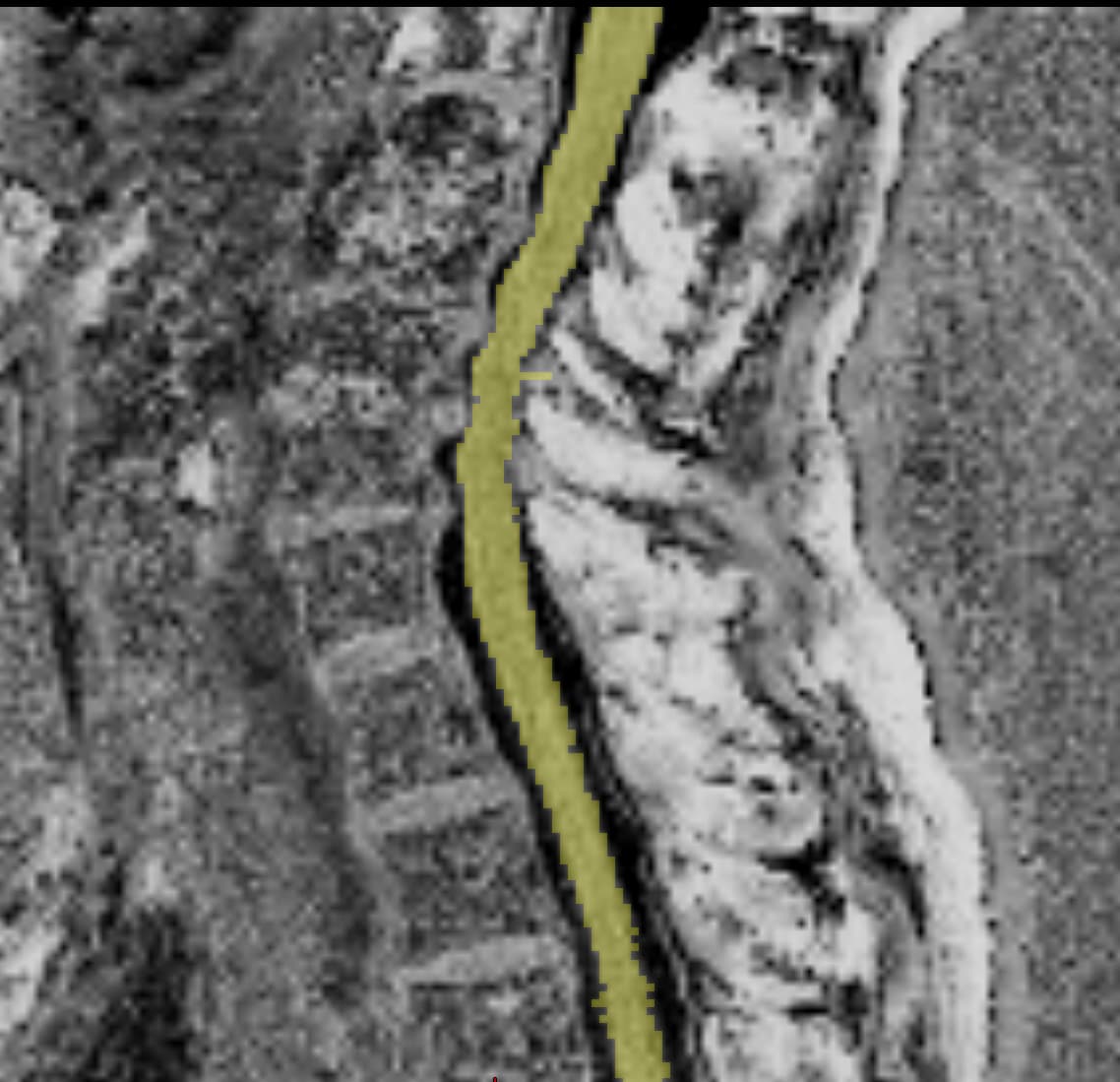
I attached a T2 image (0.8 mm isotropic) with the segmentation overlay. Voxels are missing within the mask at the level of the curvature.
The dataset that I’m using also includes T1 MP2RAGE images (1mm isotropic) with a “UNI” image that has a T1 contrast and a T1 relaxometry map that looks like a T2.
The segmentation is more accurate on these images even though some voxels are outside of the spinal cord on a few slices. I guess I will need to correct them manually. Would you recommend using these images instead of the T2.
My goal is to extract DTI metrics and MTR in the white and gray matter and in specific tracts.
Then, it might be better not to stitch the FA, MD, …maps, right?
right
The dataset that I’m using also includes T1 MP2RAGE images (1mm isotropic) with a “UNI” image that has a T1 contrast and a T1 relaxometry map that looks like a T2.
The segmentation is more accurate on these images even though some voxels are outside of the spinal cord on a few slices. I guess I will need to correct them manually. Would you recommend using these images instead of the T2.
My goal is to extract DTI metrics and MTR in the white and gray matter and in specific tracts.
If your end goal is to extract DTI metrics and MTR, then you do not need a “perfect” segmentation on the T2w or T1w. What you can do is use the T2w (or UNI, or one of the T1w with inversion pulse from the MP2RAGE scans) to do a “coarse” segmentation (feel free to use sct_smooth with a second pass to get a better segmentation without missing voxels), that is then used to get the vertebral levels with sct_label_vertebrae, and once you have the vertebral levels, you can resample them to the DWI and MT space and use sct_register_to_template with the flag -ref subject. You will also need the segmentation on the DWI space (from the moco DWI averaged, as in the tutorials) and on the MTR space (eg: from the MTon, or MToff, pick the one where the cord/CSF is best).